Step 1: make an incision
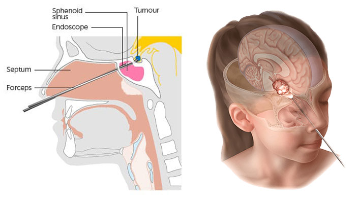
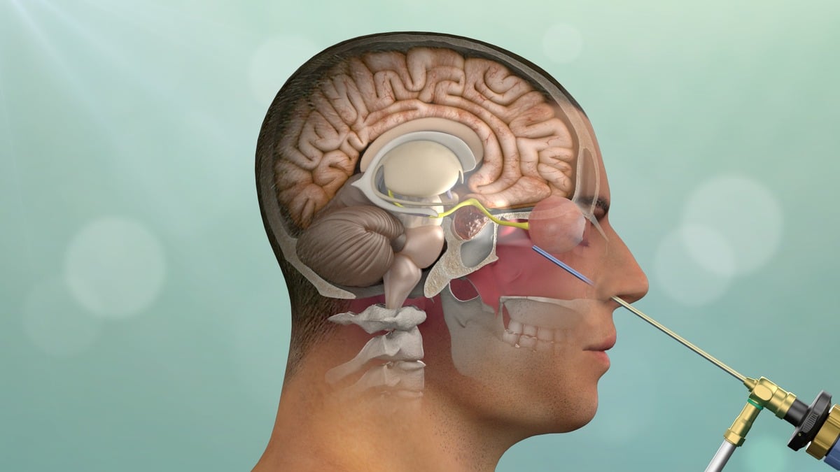
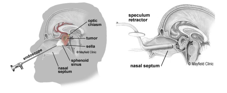
The ENT surgeon inserts the endoscope in one nostril and advances it to the back of the nasal cavity. An endoscope is a thin, tube-like instrument with a light and a camera. Video from the camera is viewed on a monitor. The surgeon passes long instruments through the nostril while watching the monitor. A small portion of the nasal septum dividing the left and right nostril is removed. Using bone-biting instruments, the front wall of the sphenoid sinus is opened (Fig. 3).
Figure 3. The endoscope is inserted through one nostril. A bony opening is made in the nasal septum (dotted line) and sphenoid sinus (green) to access the sella.
Step 2: open the sella
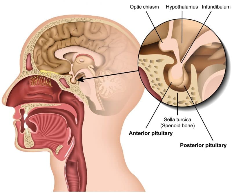
At the back wall of the sphenoid sinus is the bone overlying the pituitary gland, called the sella. The thin bone of the sella is removed to expose the tough lining of the skull called the dura. The dura is opened to expose the tumor and pituitary gland.
Step 3: remove the tumor
Through a small hole in the sella, the tumor is removed by the neurosurgeon in pieces with long grasping instruments (Fig. 4).
Figure 4. The surgeon passes instruments through the other nostril to remove the tumor.
The center of the tumor is cored out, allowing the tumor margins to fall inward so the surgeon can reach it. After all visible tumor is removed, the surgeon advances the endoscope into the sella to look and inspect for hidden tumor. Some tumors grow sideways into the cavernous sinus, a collection of veins. It may be difficult to completely remove this portion of the tumor without causing injury to the nerves and vessels. Any tumor left behind may be treated later with radiation.
Step 4: obtain fat graft (optional)
After tumor is removed, the surgeon prepares to close the sella opening. If needed, a small (2cm) skin incision is made in the abdomen to obtain a small piece of fat. The fat graft is used to fill the empty space left by the tumor removal. The abdominal incision is closed with sutures.
Step 5: close the sella opening
The hole in the sella floor is replaced with bone graft from the septum (Fig. 5). Synthetic graft material is sometimes used when there is no suitable piece of septum or the patient has had previous surgery. Biologic glue is applied over the graft in the sphenoid sinus. This glue allows healing and prevents leaking of cerebrospinal fluid (CSF) from the brain into the sinus and nasal cavity.
Figure 5. A fat graft is placed in the area where the tumor was removed. A cartilage graft is placed to close the hole in the sella. Biologic glue is applied over the area.
Soft, flexible splints may be placed in the nose along the septum to control bleeding and prevent swelling. The splints also prevent adhesions from forming that may lead to chronic nasal congestion.


